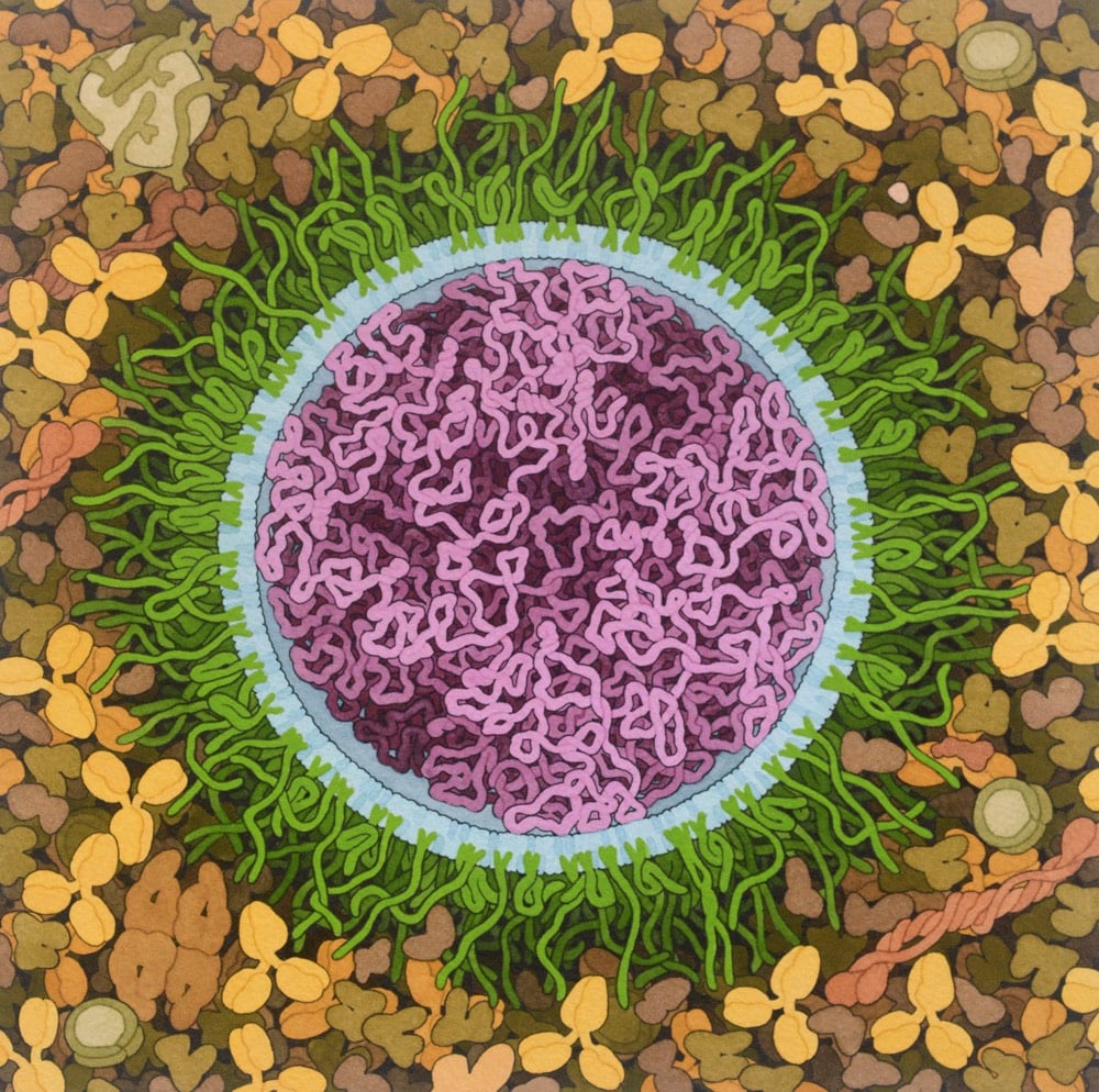
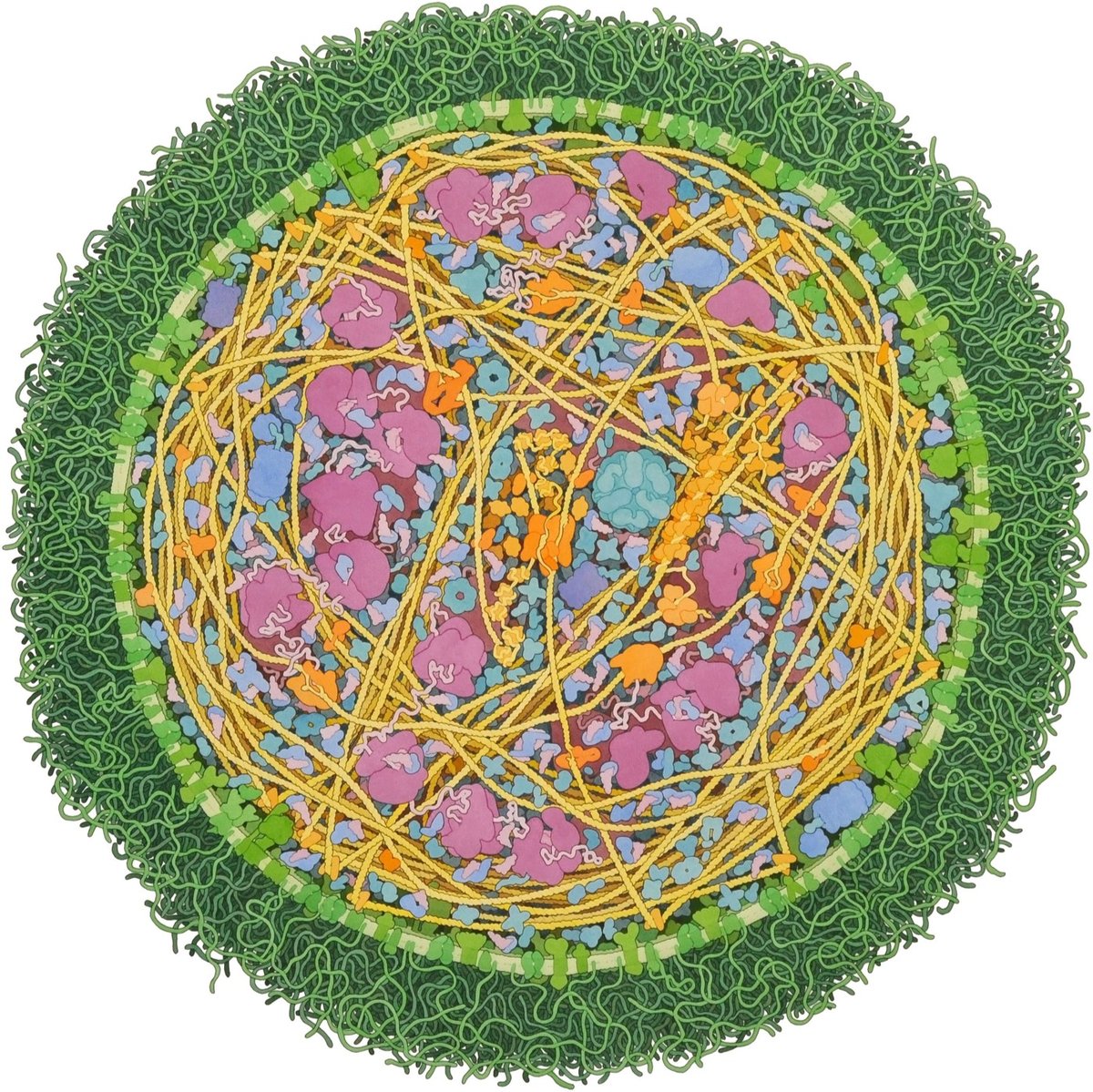
Artist and biologist David Goodsell has done a painting of the Covid-19 mRNA vaccine.
The vaccine structure is highly idealized, with spike mRNA in magenta, lipids in blue, and PEG-lipid in green. The background is blood serum or lymph.
Both the Pfizer/BioNTech and the Moderna Covid-19 vaccines are based on mRNA — you can brush up on how they work at Stat or the CDC.
mRNA vaccines are a new type of vaccine to protect against infectious diseases. To trigger an immune response, many vaccines put a weakened or inactivated germ into our bodies. Not mRNA vaccines. Instead, they teach our cells how to make a protein — or even just a piece of a protein — that triggers an immune response inside our bodies. That immune response, which produces antibodies, is what protects us from getting infected if the real virus enters our bodies.
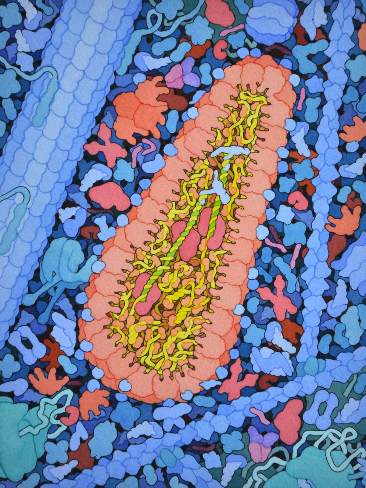
See also Goodsell’s painting of a SARS coronavirus from back in February.
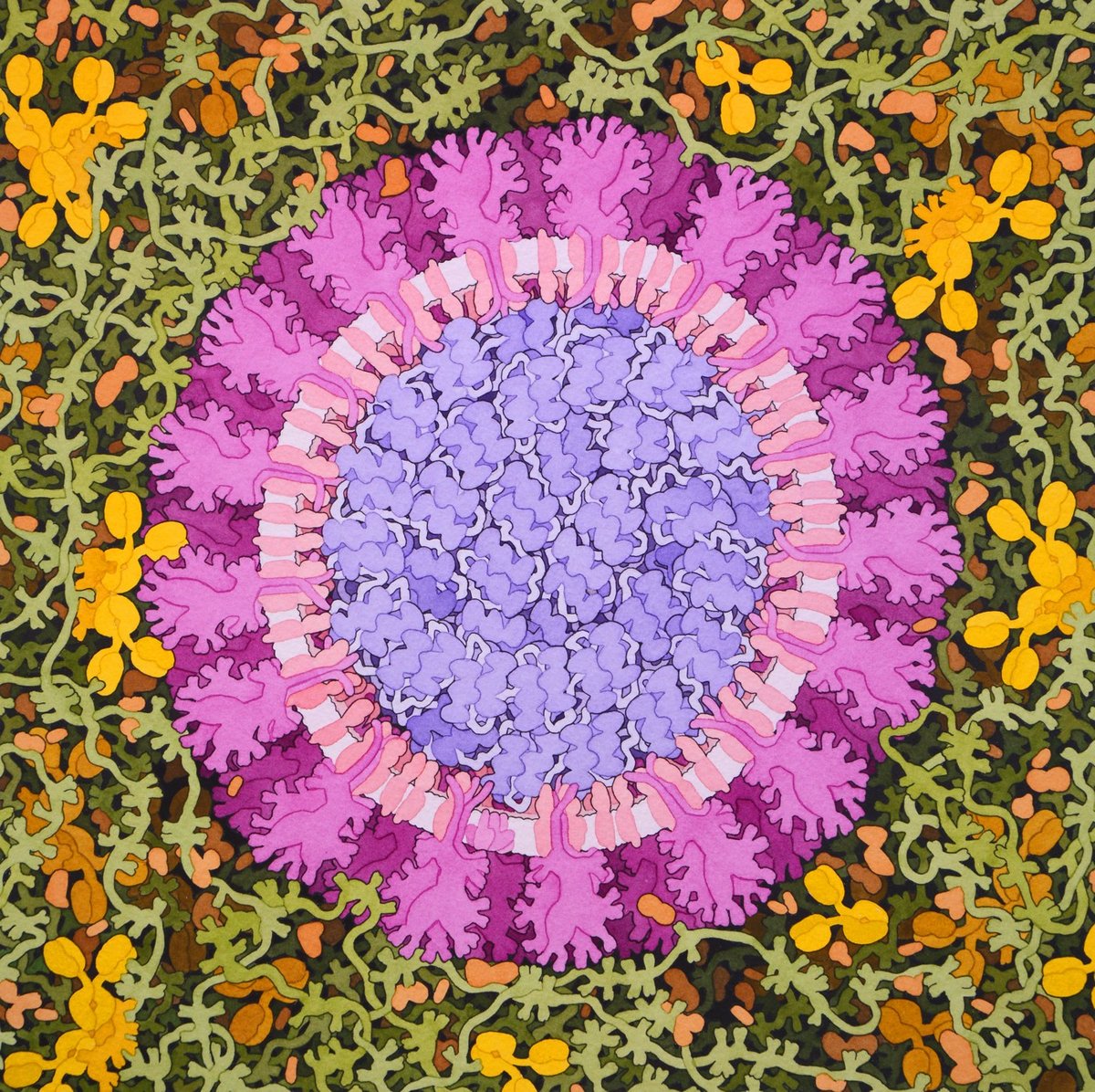
Since the early 90s, biologist David Goodsell (previously) has been creating scientifically accurate paintings of the structures of cells, molecules, and, yes, viruses. In early February, Goodsell completed a painting of a SARS coronavirus (above).
This painting depicts a coronavirus just entering the lungs, surrounded by mucus secreted by respiratory cells, secreted antibodies, and several small immune systems proteins. The virus is enclosed by a membrane that includes the S (spike) protein, which will mediate attachment and entry into cells, M (membrane) protein, which is involved in organization of the nucleoprotein inside, and E (envelope) protein, which is a membrane channel involved in budding of the virus and may be incorporated into the virion during that process. The nucleoprotein inside includes many copies of the N (nucleocapsid) protein bound to the genomic RNA.
In a brief interview with the NY Times, Goodsell explained why he made the image:
“You have to admit, these viruses are so symmetrical that they’re beautiful,” said Mr. Goodsell, an associate professor at Scripps Research Institute in La Jolla. “Are bright colors and pretty stuff the right approach? The jury’s still out. I’m not trying to make these things look dangerous, I want people to understand how they’re built.”
Seeing the infection count rise, Mr. Goodsell said he worried about the health of his aging parents in Los Angeles. But he hopes his painting can quell fears about the novel coronavirus by educating people on the virus’s workings: “I want people to think of viruses as being an entity that we can learn about and fight. They’re not nebulous nothings.”
Goodsell is currently working on a painting featuring the life cycle of a coronavirus and sharing his progress on Twitter. (via @christopherjobs)
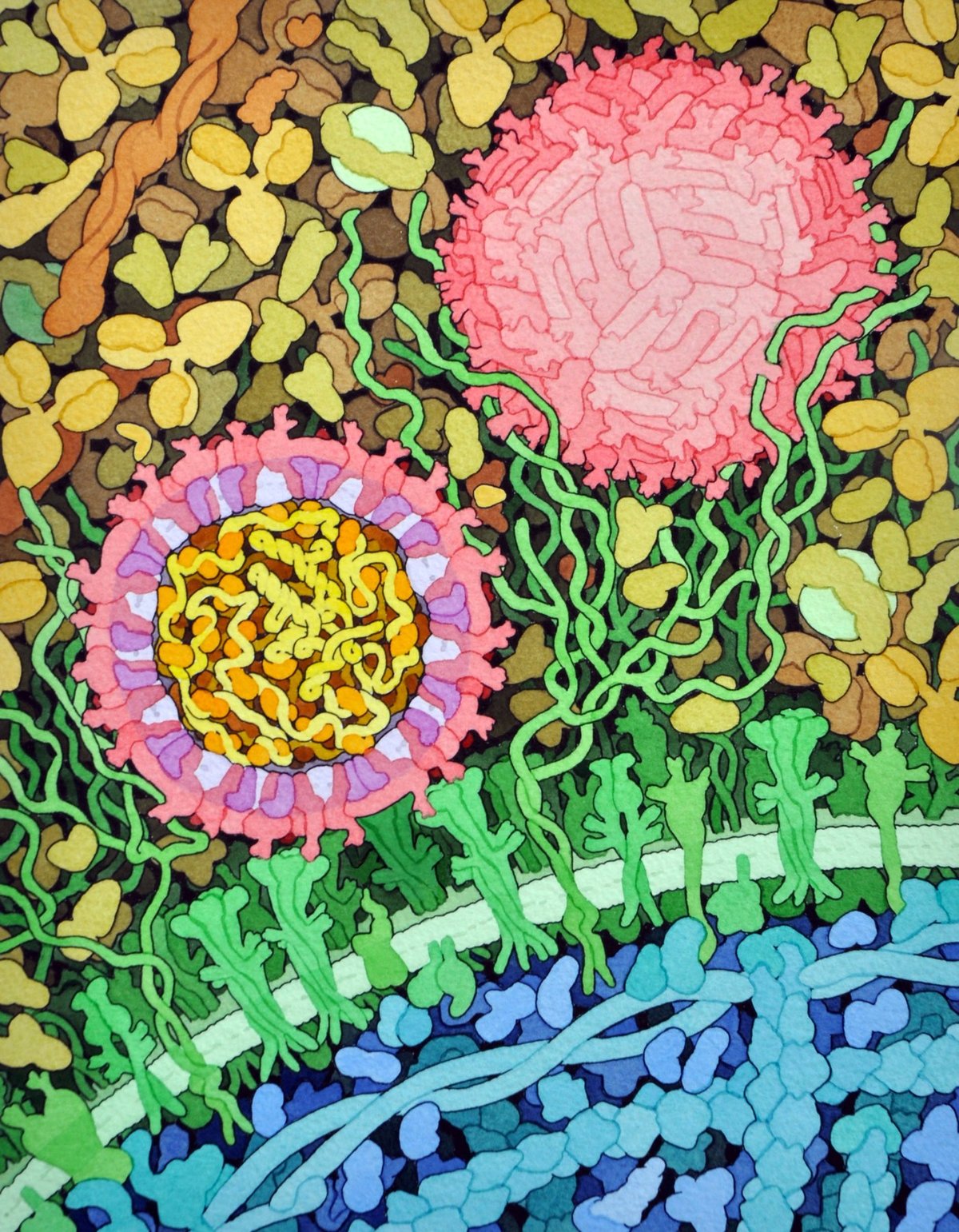
For more than 25 years, biologist David Goodsell has been making scientifically accurate paintings and illustrations of the molecular structures of things related to HIV, cancer cells, ebola, Zika, diabetes, proteins, enzymes, and hundreds of other scientific and medical processes.
Since the early 1990s, I have been working with a type of illustration that shows portions of living cells magnified so that you can see individual molecules. I try to make these illustrations as accurate as possible, using information from atomic structure analysis, electron microscopy, and biochemical analysis to get the proper number of molecules, in the proper place, and with the proper size and shape.
Much of his work is available to use for free (with attribution) and is scattered across the web: the Molecule of the Month, Molecular Landscapes, Illustrations for Public Use. He has also published several books of his paintings, the most popular of which is The Machinery of Life. Science magazine recently profiled Goodsell and his work.
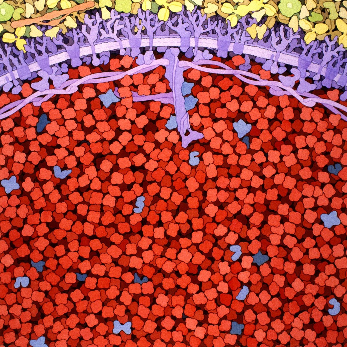
In addition to studying pictures of cells from high-powered microscopes, Goodsell relies on molecular structures from electron microscopy (EM), x-ray crystallography, and nuclear magnetic resonance spectroscopy to make his paintings, which show the often crowded and complex world of cells and the microbes that infect them. He even uses the known weights of molecules if that’s all he has so that he can at least draw, say, a correctly sized circle. “I’m a scientist first,” he says. “I’m not making editorial images that are meant to sell magazines. I want to somehow inform the scientists and armchair scientists what the state of knowledge is now and hopefully give them an intuitive sense of how these things really look — or may look,” he says.
May look?
“These pictures have tons and tons and tons of artistic license,” he says. “They’re just one snapshot of something that’s intrinsically superdynamic. Every time I do a painting, the next day it’s out of date because there’s so much more data coming out.”
Here’s a quick video profile as well:
All images are by David S. Goodsell, the Scripps Research Institute. (via alexandra kammen)
Socials & More